Last updated: July 2026 • 8 min read
Quick Answer
Western blot interpretation requires more than comparing band intensity. It involves evaluating whether the experimental evidence supports the authors’ conclusions.
Reading a Western blot figure isn’t about deciding whether one band is darker than another.
It’s about asking whether the experimental evidence actually supports the authors’ conclusion.
A careful interpretation starts with the scientific claim, then moves through the bands, the controls, the quantitative data, and finally the biological interpretation.
A Western blot is evidence—not the conclusion itself.
Why Western Blot Figures Are Often Misunderstood
Most researchers learn how to perform a Western blot.
Far fewer learn how to evaluate one.
Those are very different skills.
When reading a paper, it’s easy to focus on the image itself.
One lane looks darker. Another looks weaker.
At first glance, the conclusion seems obvious.
But a Western blot figure is only one part of the evidence.
Before deciding whether it supports the authors’ claim, it’s worth asking a few questions.
- What was the experiment designed to test?
- Were the controls appropriate?
- Was the result reproduced consistently?
Those questions usually tell you much more than the bands alone.
This guide explains the workflow I use to evaluate Western blot figures in scientific papers.
Western Blot Interpretation Workflow
One of the easiest mistakes to make is judging the blot before understanding the experiment.
I try to do the opposite.
The scientific claim always comes first.
Everything else follows from there.
A typical workflow looks like this:
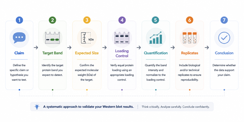
| Step | Question |
|---|---|
| Scientific claim | What are the authors trying to demonstrate? |
| Target band | Is the correct protein being detected? |
| Expected size | Does the band appear at the expected molecular weight? |
| Loading control | Were similar amounts of protein loaded? |
| Quantification | Do the measurements support the visual impression? |
| Replicates | Was the result reproduced consistently? |
| Conclusion | Does the evidence justify the authors’ interpretation? |
Notice where the workflow begins.
Not with the image.
With the scientific question the figure is meant to answer.
Western Blot Interpretation
Step 1: Start With the Scientific Claim
Before examining the blot itself, start with a simple question:
What are the authors actually trying to prove?
In most papers, the claim falls into one of a few categories.
- Protein expression increased or decreased
- Protein phosphorylation changed
- A signaling pathway was activated
- Gene knockdown reduced protein abundance
- A treatment altered protein levels
Once the claim is clear, the figure becomes much easier to interpret.
Without that context, it’s easy to focus on details that have little relevance to the experiment.
Never evaluate a blot before knowing what it is trying to prove.
Step 2: Look at the Bands
Only after understanding the scientific question is it time to examine the blot itself.
Rather than asking whether one band is darker than another, start with a simple checklist.
Before interpreting the band, check:

✔ Expected molecular weight

✔ Antibody specificity

✔ Unexpected bands

✔ Signal quality
Check the following:
- Does the band appear at the expected molecular weight?
- Is there evidence of non-specific binding?
- Are unexpected bands present?
- Is the signal strong enough to interpret confidently?
A darker band can suggest increased protein abundance, but only if the antibody is specific, the exposure is appropriate, and the experiment has been properly controlled.
Without that context, band intensity alone can be misleading.
The most convincing Western blot isn’t always the cleanest one.
It’s the one that makes biological sense.
Step 3: Check the Controls
After evaluating the target protein, move directly to the controls.
Loading controls such as β-actin, GAPDH, α-tubulin, or validated total protein stains help determine whether similar amounts of protein were loaded into each lane.
They provide context for interpreting differences in band intensity.
What they do not do is prove that the experiment itself is correct.
A loading control cannot confirm antibody specificity or validate the biological interpretation.
It answers one experimental question:
Were the samples loaded consistently?
A clean loading control strengthens the interpretation.
It does not prove it.
Step 4: Look for Quantification
A representative blot tells only part of the story.
Quantitative analysis provides the context needed to judge how convincing the result really is.
Before accepting the conclusion, look for:

✔ Quantification

✔ Biological replicates

✔ Statistical analysis

✔ Consistent trends
Look for:
- Densitometric quantification
- Biological replicates
- Appropriate statistical analysis
- Consistent trends across independent experiments
A dramatic-looking band does not always correspond to a strong experimental result.
Conversely, a modest visual difference may become convincing when supported by reproducible quantitative data.
The image shows the evidence.
Quantification tells you how much confidence that evidence deserves.
Real Paper Example
Let’s imagine you’re reading a paper claiming that AKT phosphorylation increases after treatment.
The figure looks something like this:
p-AKT Band
      ↓
Total AKT
      ↓
Loading Control
      ↓
Quantification
      ↓
Interpretation
At first glance, the treated sample shows a stronger p-AKT band.
That alone is not enough to support the conclusion.
Before accepting the interpretation, check:
- Is the phosphorylated band at the expected molecular weight?
- Is total AKT relatively unchanged?
- Does the loading control look consistent?
- Does the quantified data support what the eye sees?
- Was the result reproduced across independent experiments?
Only after those questions have been answered is it reasonable to interpret the biological significance of the result.
A Western blot rarely stands on its own.
It becomes convincing when the surrounding evidence supports it.
Common Interpretation Mistakes
The biggest mistake isn’t misunderstanding the image. It’s drawing conclusions before evaluating the evidence.
❌ “A darker band means more protein.”
Why it’s a problem
Not necessarily. Band intensity can be influenced by exposure conditions, antibody performance, protein loading, and image processing.
Better approach
Evaluate band intensity only after considering the controls, quantification, and experimental context.
❌ “A loading control proves the experiment is reliable.”
Why it’s a problem
Loading controls only demonstrate that similar amounts of protein were loaded into each lane.
They do not validate antibody specificity or biological interpretation.
Better approach
Treat loading controls as one piece of evidence—not the final proof.
❌ “One representative blot is enough.”
Why it’s a problem
A single image cannot demonstrate reproducibility. Representative blots should be supported by biological replicates and quantitative analysis.
Better approach
Look for replicated experiments, statistical analysis, and consistent results before accepting the conclusion.
❌ “Strong exposure always = more protein”
Why it’s a problem
A stronger signal may simply reflect longer exposure rather than higher protein abundance.
Better approach
Always interpret band intensity together with exposure conditions, controls, and quantitative analysis.
❌ “A cropped blot is always acceptable”
Why it’s a problem
Cropping is common in scientific figures. However, excessive cropping can hide non-specific bands, image artifacts, or important experimental context.
Better approach
When available, examine uncropped blots or supplementary figures rather than relying solely on the cropped image.
❌ “Clean blot = good experiment”
Why it’s a problem
A visually clean blot can still suffer from poor antibody specificity, inadequate controls, or limited biological replication.
Better approach
Judge the experiment—not the aesthetics of the image.
What Should You Check First?
Whenever I evaluate a Western blot figure, I come back to the same checklist.
| Check | Why It Matters |
|---|---|
| Scientific claim | Understand what the experiment is trying to demonstrate |
| Band size | Confirm the correct protein is being detected |
| Loading control | Check whether protein loading is comparable |
| Quantification | Determine whether the visual impression is supported by measurements |
| Replicates | Assess whether the findings are reproducible |
These five questions prevent many of the most common interpretation mistakes.
Just as importantly, they encourage you to evaluate the experiment before interpreting the image.
Key Takeaways
- Start with the scientific claim, not the image.
- A band should always be interpreted in context.
- Loading controls support interpretation—they do not prove it.
- Quantification and biological replicates strengthen confidence in the result.
- A Western blot figure should be evaluated as part of the evidence, not as the conclusion itself.
Frequently Asked Questions
Q1. How do you read a Western blot figure?
Start with the scientific claim, then evaluate the target band, loading control, quantification, and biological replicates before interpreting the conclusion.
Q2. Does a darker band always mean more protein?
No.
Band intensity can be affected by several experimental factors and should always be interpreted alongside appropriate controls and quantitative analysis.
Q3. Why are loading controls important?
Loading controls help determine whether comparable amounts of protein were loaded across samples.
They provide context for comparing band intensity.
Q4. Is one Western blot image enough?
Usually not.
Representative images should be supported by replicate experiments and quantitative data.
Q5. What should I check before trusting a Western blot figure?
At a minimum:
- The scientific claim
- The expected band size
- The loading control
- Quantitative analysis
- Biological replicates
Q6. Can Western blot prove protein expression changes?
A Western blot can support that conclusion.
It rarely proves it on its own.
Interpretation should always consider the broader experimental design and supporting evidence.
Final Thoughts
A Western blot band is evidence. It is never the conclusion by itself.
Further Reading
- Abcam – Western blot guide
- Bio-Rad Laboratories – Western blot handbook
- Cell Signaling Technology – Western blot protocol