Last updated: July 2026
Your target band is there—but so is everything else.
Instead of a clean membrane, the entire blot appears gray, covered with speckles, or filled with unwanted bands. The signal may be strong, but so is the background, making it difficult to interpret the result with confidence.
A common reaction is to increase washing, dilute the primary antibody, or try a different blocking buffer. Sometimes these changes help. Just as often, they do not—because not every background problem has the same cause.
The first step is not changing the protocol.
It’s identifying what kind of background you’re looking at.
Experienced researchers rarely troubleshoot high background by randomly adjusting antibody concentrations. Instead, they examine the membrane itself. A uniformly gray membrane, scattered speckles, patchy staining, and multiple nonspecific bands each point toward different underlying problems.
In this guide, you’ll learn how to recognize the most common background patterns, understand what usually causes them, and troubleshoot them in a logical order.
Key principle
Don’t treat every background problem the same. Identify the pattern first, then adjust the protocol.
🚨 High Background? Start Here
Not all background is caused by the same problem. The appearance of your membrane is often the fastest clue to what went wrong.
If you’re not sure where to begin, work through this checklist before changing multiple protocol conditions.
| ✓ | Check | If Background Persists |
|---|---|---|
| □ | Shorten the exposure time | Dilute the secondary antibody |
| □ | Prepare fresh TBST | Increase washing |
| □ | Use an appropriate blocking reagent | Try milk or BSA if appropriate |
| □ | Optimize the secondary antibody | Reduce the primary antibody concentration |
| □ | Match ECL sensitivity to your target | Consider a lower-sensitivity substrate |
Most background problems can be narrowed down within a few minutes using this checklist.
Identify the Pattern
Before changing your protocol, spend a few seconds looking carefully at the membrane.
The appearance of the background is often your biggest clue.
Different background patterns usually point to different technical problems. Identifying the pattern first helps narrow the list of possible causes before you begin changing the protocol.
| What you see | Most likely cause | Try this first |
|---|---|---|
| Uniform gray background | Excessive antibody concentration or overexposure | Shorten exposure, then optimize the secondary antibody |
| Random speckles | Contaminated buffers or antibody precipitates | Prepare fresh buffers and clean the equipment |
| Patchy background | Uneven blocking or membrane drying | Keep the membrane wet and improve reagent coverage |
| Multiple nonspecific bands | Antibody cross-reactivity | Reduce the primary antibody concentration |
| Strong target, high background | Poor signal-to-noise ratio | Reduce exposure before changing antibodies |
Think of this as a starting point rather than a definitive diagnosis. More than one factor may contribute to high background, but identifying the pattern is often the fastest way to narrow down the likely causes.
💡 Lab Tip
Before changing your protocol, identify the background pattern first. It often narrows the list of possible causes more effectively than adjusting antibodies by trial and error.
Your Entire Membrane Looks Gray
A uniformly gray membrane is one of the most common forms of Western blot background.
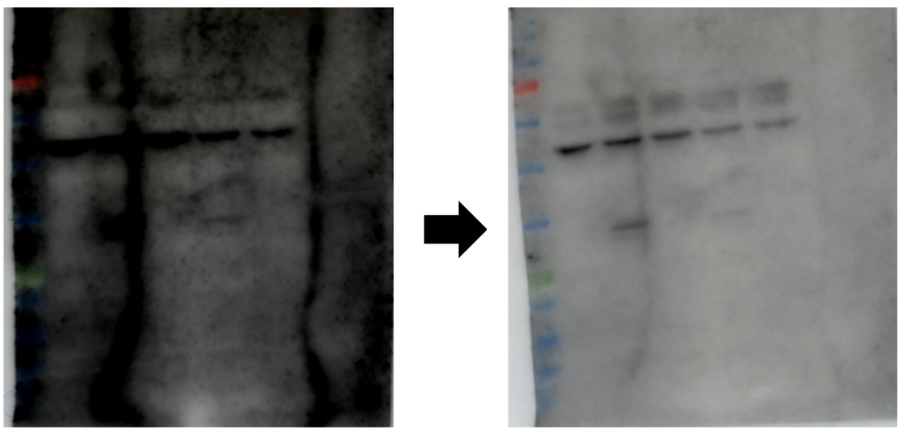
What This Usually Means
The overall signal is being amplified rather than the target band alone.
What Usually Causes It?
Uniform background is most commonly associated with excessive nonspecific signal rather than a localized experimental error.
Common causes include:
- Primary antibody concentration is too high.
- Secondary antibody concentration is too high.
- Exposure time is too long.
- The ECL substrate is more sensitive than necessary for the target protein.
What Should You Try First?
Rather than changing several conditions simultaneously, optimize one variable at a time.
A practical troubleshooting order is:
- Reduce the exposure time.
- Dilute the secondary antibody.
- If needed, optimize the primary antibody concentration.
- Consider using a less sensitive ECL substrate if your target protein is highly abundant.
This sequence minimizes unnecessary optimization while preserving specific signal whenever possible.
💡 Lab Tip
Optimize the secondary antibody first. It is often responsible for widespread background and is usually easier to adjust than the primary antibody.
You’re Seeing Random Speckles
Speckles appear as scattered dark spots or irregular dots across the membrane.
What This Usually Means
The problem is usually physical contamination rather than antibody specificity.
Dust, dirty forceps, old buffers, or antibody precipitates are all common causes.
Common Causes
Possible causes include:
- Antibody precipitates
- Dust particles
- Dirty forceps
- Contaminated incubation trays
- Old or contaminated buffers
Because these artifacts are physical rather than biological, they often appear randomly across the membrane instead of following the protein migration pattern.
Quick Fixes
Before changing antibody concentrations, check the simplest possibilities first.
Start with the simplest possibilities before changing antibody concentrations.
- Prepare fresh wash and incubation buffers.
- Use clean containers and forceps.
- Prepare a fresh antibody solution if precipitation is suspected.
- Consult the manufacturer’s recommendations before filtering antibodies that are prone to precipitation.
If these simple changes eliminate the speckles, further antibody optimization is usually unnecessary.
You’re Seeing Patchy Background
Patchy background appears as irregular regions of high and low signal across the membrane.
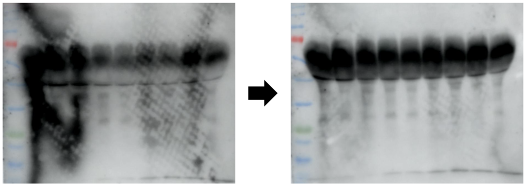
This pattern usually indicates that different parts of the membrane were exposed to different experimental conditions.
What This Usually Means
Different parts of the membrane were exposed to different conditions.
Uneven blocking, incomplete reagent coverage, or membrane drying are common explanations.
Common Causes
Typical causes include:
- Membrane drying during the protocol
- Uneven blocking
- Incomplete coverage with antibody or wash buffer
- Insufficient agitation during incubation
Practical Fixes
The solution is usually to improve reagent coverage and prevent the membrane from drying.
- Use enough blocking solution to completely cover the membrane.
- Gently agitate the membrane during blocking and antibody incubation.
- Ensure the membrane remains fully submerged throughout the experiment.
- Never allow the membrane to dry between protocol steps.
Small differences in reagent coverage can produce surprisingly large differences in background.
You See Multiple Unexpected Bands
Sometimes the problem is not a gray membrane but numerous unexpected bands.
In this situation, the target protein may still be visible, but interpretation becomes difficult because additional bands appear at multiple molecular weights.
What This Usually Means
The antibody is binding to proteins other than your target.
This is usually caused by cross-reactivity or suboptimal antibody conditions rather than transfer or detection problems.
Common Causes
Multiple nonspecific bands are most commonly associated with:
- Primary antibody cross-reactivity
- Excessive antibody concentration
- Sample-specific protein interactions
What Should You Optimize?
Start by optimizing antibody specificity rather than changing the detection system.
A practical order is:
- Reduce the primary antibody concentration.
- Optimize antibody incubation conditions.
- Review whether the antibody has been validated for Western blotting and for your sample type.
- If available, compare the pattern with a positive control.
Optimize one condition at a time so you can identify which change actually improves specificity.
Your Target Band Is Strong, but So Is the Background
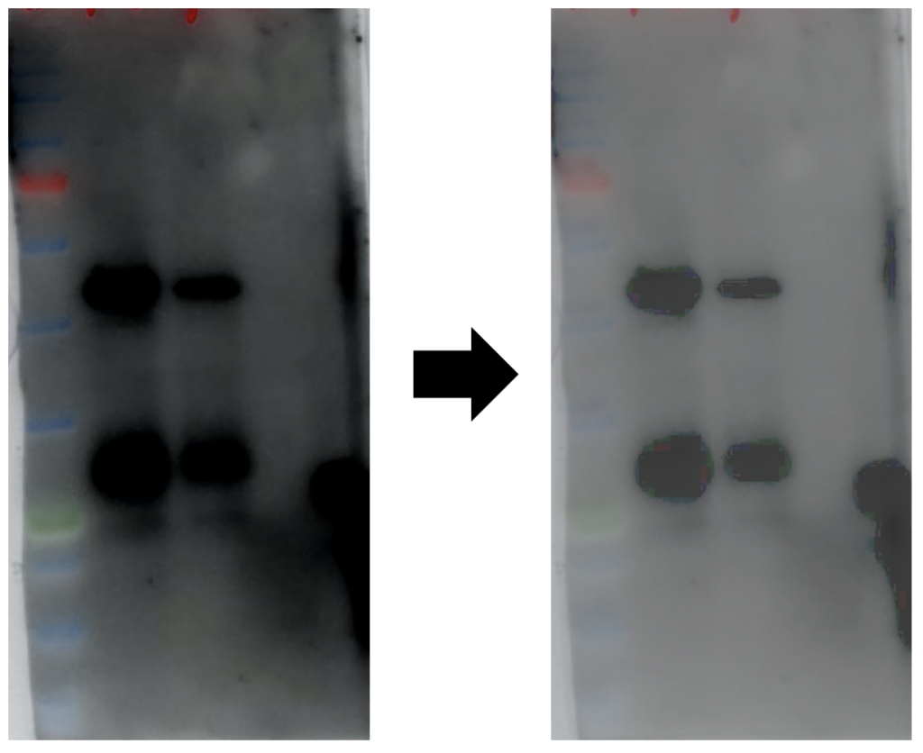
If your target band is already strong, don’t try to make it stronger.
Instead, improve the signal-to-noise ratio.
When the target band is already obvious, increasing signal is rarely the answer. Instead, improve the signal-to-noise ratio by making one change at a time:
- Shorten the exposure time.
- Increase washing if background remains high.
- Dilute the secondary antibody.
- If your target is highly abundant, consider using a less sensitive ECL substrate.
💡 Lab Tip
If the target band is already obvious, adding more primary antibody usually increases background rather than improving the blot.
Blocking and Washing Considerations
Blocking and washing are often treated as routine protocol steps, but together they determine how much nonspecific antibody remains on the membrane.
Small improvements here can dramatically reduce background without changing antibodies.
Milk vs. BSA
Both milk and BSA are widely used blocking reagents, but they are not interchangeable in every experiment.
Nonfat dry milk
- Cost-effective and commonly used.
- Suitable for many routine Western blot applications.
BSA
- Often preferred for phospho-specific antibodies because milk contains casein, a phosphoprotein that may interfere with phospho-antibody detection.
For many routine Western blots, either blocker performs well. However, blocking recommendations vary among antibodies, so always consult the antibody datasheet for your specific application.
How Much Washing Is Enough?
There is no universal washing protocol that works for every Western blot.
Instead of immediately changing antibody concentrations, first consider whether washing conditions are adequate.
Good practice includes:
- Increasing wash duration before changing antibody dilution.
- Using enough wash buffer to completely cover the membrane.
- Gently agitating the membrane throughout each wash.
- Preparing fresh TBST when contamination is suspected.
Rather than making large changes all at once, adjust one washing parameter at a time and evaluate its effect on background.
Before Repeating Your Western Blot
Before repeating the entire experiment, confirm that the problem cannot be corrected by adjusting the detection conditions or membrane handling.
| ✓ | Quick check | Why it helps |
|---|---|---|
| □ | Confirm that the secondary antibody matches the primary antibody. | A species mismatch can increase background or eliminate signal. |
| □ | Prepare fresh ECL before developing the membrane again. | Fresh substrate can improve signal quality and rule out detection-related problems. |
| □ | Probe a housekeeping protein if you’re unsure whether the problem is target-specific. | A normal housekeeping band suggests the workflow is working and the issue is likely target-specific. |
| □ | Increase exposure gradually instead of jumping to the longest exposure. | Longer exposure also amplifies background. |
| □ | If only one molecular weight is relevant, consider cutting the membrane before antibody incubation. | This may reduce unnecessary background and antibody consumption when the target molecular weight is well established. |
| □ | If protein loading and transfer were successful, consider stripping and reprobing the membrane before repeating the experiment. | This can help distinguish technical problems from target-specific issues. |
Common Troubleshooting Mistakes
Increasing the Primary Antibody First
Many researchers immediately increase the primary antibody concentration when background appears.
In reality, this often increases nonspecific binding before improving the target signal.
Optimize the secondary antibody first whenever possible.
Assuming More Washing Solves Everything
Additional washing can reduce residual antibody, but it cannot compensate for excessive antibody concentration, poor blocking, or overexposure.
Treat washing as one part of the troubleshooting process—not the universal solution.
Letting the Membrane Dry
Dry membranes frequently develop uneven or patchy background that cannot easily be corrected.
Keep the membrane completely submerged throughout blocking, antibody incubation, and washing.
Changing Multiple Variables at Once
Reducing antibody concentration, changing blocking buffer, increasing washing, and switching ECL simultaneously may improve the blot—but you will not know which change actually solved the problem.
Optimize one variable at a time whenever practical.
Frequently Asked Questions
Q1. Why is my Western blot background so high?
High background is usually caused by nonspecific antibody binding, insufficient washing, excessive exposure, or an overly sensitive detection system rather than a single technical error.
Q2. Does too much antibody increase background?
Yes.
Both primary and secondary antibodies can increase nonspecific signal when used at excessively high concentrations.
Q3. Should I use milk or BSA?
Neither is universally better.
Milk performs well for many routine Western blots, whereas BSA is often preferred for phospho-specific antibodies.
Always consider the antibody manufacturer’s recommendations for your application.
Q4. Can ECL increase background?
Yes.
Highly sensitive ECL substrates can amplify weak nonspecific signal as well as the desired target band.
Q5. Can overexposure cause high background?
Yes.
Long exposure times increase both signal and background, making the membrane appear noisier than it actually is.
Q6. Should I repeat the experiment?
Not immediately.
First identify the background pattern and optimize the most likely contributing variable. Many background problems can be corrected without repeating the entire Western blot.
Q7. Why is my Western blot background only around the edges?
Edge background is usually caused by uneven reagent coverage, membrane drying, or incomplete agitation during incubation.
Key Takeaways
If your Western blot has high background, remember these principles:
- Identify the background pattern before changing the protocol.
- Optimize one variable at a time.
- Start with exposure time and the secondary antibody before adjusting the primary antibody.
- Keep the membrane fully wet throughout the experiment.
- Match ECL sensitivity to the expected abundance of your target protein.
- Systematic troubleshooting is more effective than changing multiple conditions simultaneously.
Above all, remember this:
The appearance of the background is often the biggest clue to its cause. Every background pattern tells a story. Learning to recognize those patterns will usually save more time than changing multiple protocol conditions at once.
Continue Learning
High background is only one type of Western blot troubleshooting problem.
You may also find these guides helpful:
Western Blot Troubleshooting
- Western Blot No Bands? A Step-by-Step Troubleshooting Guide
- Weak Western Blot Bands: Causes and Proven Fixes
- Western Blot Smiling Bands: Causes and Solutions
- Western Blot Multiple Bands: Causes and Solutions
Western Blot Fundamentals
- How to Read Western Blot Results
- Western Blot Controls Explained
- Loading Controls in Western Blot
- PVDF vs. Nitrocellulose Membranes
- Chemiluminescence vs. Fluorescence Detection