Last updated: July 2026
If you’ve ever opened a biology paper, chances are you’ve come across a Western blot figure—a series of dark bands lined up across a membrane.
At first glance, it can look surprisingly simple.
A few bands. Some labels. Perhaps a loading control underneath.
But behind those bands is one of the most widely used techniques in molecular biology for detecting specific proteins.
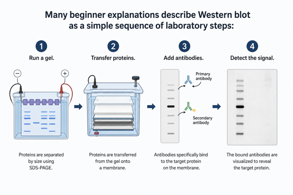
While technically correct, this approach often misses the bigger picture.
Every step in a Western blot exists for a specific reason. Understanding why each step is necessary is far more useful than memorizing the protocol itself.
Once you understand the purpose of each step, Western blot becomes much easier to interpret—and much easier to troubleshoot when something goes wrong.
This is especially important because most Western blot problems are not caused by a single mistake, but by understanding which part of the workflow failed.
In this guide, we’ll explain Western blot from the perspective of what each step accomplishes, rather than simply describing what happens in the laboratory.
What Does a Western Blot Actually Detect?
The simplest way to understand Western blot is to start with what it measures.
A Western blot detects proteins.
It does not detect DNA or RNA.
This distinction is important because different molecular biology techniques answer different biological questions.
| Technique | Detects | Best for |
|---|---|---|
| PCR | DNA | Detecting genes or DNA sequences |
| RT-qPCR | mRNA | Measuring changes in gene expression at the transcript level |
| Western blot | Protein | Detecting protein expression, size, and some post-translational modifications |
| ELISA | Protein | Measuring protein abundance across many samples |
| Immunohistochemistry | Protein | Determining where a protein is located within tissue |
For example:
- If you want to know whether a gene is present, PCR is the appropriate technique.
- If you want to measure gene expression, RT-qPCR measures mRNA.
- If you want to determine whether the protein itself is present—and whether it appears at the expected molecular weight—a Western blot is often the better choice.
Measuring mRNA does not necessarily tell you whether the corresponding protein is present.
Why Do Researchers Use Western Blot?
Western blot is widely used because it answers questions that many other techniques cannot.
Rather than measuring DNA or RNA, it allows researchers to detect a specific protein within a complex mixture containing thousands of different proteins.
Common applications include:
- Comparing protein expression between experimental groups
- Confirming successful gene knockout or knockdown
- Validating CRISPR-mediated gene editing at the protein level
- Measuring activation of signaling pathways using phospho-specific antibodies
- Confirming overexpression of recombinant proteins
In practice, researchers often use Western blot to answer questions such as:
Did the treatment increase protein expression?
Is the knockout protein truly absent?
Was the signaling pathway activated after stimulation?
Notice that each question focuses on protein-level evidence.
That is the strength of Western blot.
This combination of specificity and molecular weight information is one of the main reasons Western blot remains widely used in molecular and cellular biology.
How Does Western Blot Work?
At its core, Western blot is a step-by-step workflow that gradually narrows thousands of proteins down to one specific target.
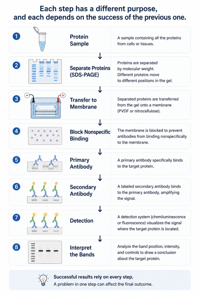
The most useful way to learn Western blot is to understand the purpose behind each step.
| Step | Purpose |
|---|---|
| SDS-PAGE | Separate proteins according to molecular weight. |
| Transfer | Move proteins from the gel onto a membrane for antibody detection. |
| Blocking | Prevent antibodies from binding nonspecifically to the membrane. |
| Primary antibody | Bind specifically to the target protein. |
| Secondary antibody | Bind the primary antibody and amplify the detection signal. |
| Detection | Produce a visible signal where the target protein is located. |
| Interpretation | Decide whether the observed bands support the biological conclusion. |
Every step contributes to a single goal: identifying one specific protein within a complex mixture.
Why Is Each Step Necessary?
Understanding the purpose of each step makes Western blot much easier to learn—and much easier to troubleshoot.
Step 1 — Separate Proteins
Purpose: Separate proteins by molecular weight.
A cell contains thousands of different proteins. If they were detected without first being separated, their signals would overlap and become impossible to interpret.
SDS-PAGE solves this problem by separating proteins according to their size before antibody detection begins.
Step 2 — Transfer to a Membrane
Purpose: Move proteins onto a stable surface that antibodies can access.
Proteins remain trapped inside the polyacrylamide gel after electrophoresis.
Transferring them onto a membrane—typically PVDF or nitrocellulose—creates a durable surface where antibodies can bind efficiently during the following steps.
Step 3 — Block the Membrane
Purpose: Prevent antibodies from sticking everywhere.
The membrane contains many sites where antibodies could bind nonspecifically.
Blocking fills these potential binding sites with proteins that reduce nonspecific antibody attachment, lowering background signal.
Step 4 — Primary Antibody
Purpose: Recognize the target protein.
The primary antibody is responsible for specificity.
It binds only to the protein—or protein modification—it was designed to recognize, allowing researchers to distinguish the target from thousands of other proteins present on the membrane.
Step 5 — Secondary Antibody
Purpose: Generate a detectable signal.
The secondary antibody binds to the primary antibody rather than the target protein itself.
Because multiple secondary antibodies can bind to a single primary antibody, this step amplifies the signal and makes low-abundance proteins much easier to detect.
Step 6 — Detection
Purpose: Visualize where the target protein is located.
After antibody binding, the signal is converted into a visible image.
The two most common detection methods are:
- Chemiluminescence, which produces light through an enzyme-substrate reaction.
- Fluorescence, which detects fluorescently labeled antibodies using an imaging system.
Regardless of the detection method, the final result is the same: a band that represents the location of the target protein on the membrane.
The next challenge is interpreting what those bands actually mean.
Want to learn how to interpret the final bands?
→ Read: How to Read Western Blot Figures
What Do the Bands Mean?
The final image is often the first thing people notice in a Western blot.
But a band is more than a dark line on a membrane.
Its position, appearance, and intensity all provide different types of information.
| Band feature | What it tells you |
|---|---|
| Position | Approximate protein size (molecular weight) |
| Intensity | Relative signal strength (when within the linear detection range) |
| Missing band | No detectable protein or a technical problem |
| Multiple bands | Protein isoforms, post-translational modifications, or nonspecific antibody binding |
One of the most common beginner mistakes is assuming that a darker band automatically means more protein.
It doesn’t.
Band intensity is influenced by many factors, including protein loading, transfer efficiency, antibody performance, exposure time, and signal saturation.
A Western blot should always be interpreted together with appropriate controls—not by band intensity alone.
💡 Lab Tip
A darker band is not automatically a stronger result. Always ask whether the band falls within the linear detection range and whether the controls support your interpretation.
Common Beginner Misunderstandings
Many misunderstandings about Western blot arise because the final image looks deceptively simple.
Here are some of the most common.
Western Blot Measures Protein—Not Gene Expression
Western blot measures protein abundance.
If your question is about mRNA expression, techniques such as RT-qPCR are more appropriate.
A Darker Band Doesn’t Always Mean More Protein
Band intensity is influenced by protein loading, transfer efficiency, antibody performance, and exposure conditions—not only by protein abundance.
Western Blot Is Semi-Quantitative
Western blot is commonly used to compare relative protein expression between samples.
However, reliable quantification requires appropriate normalization and careful control of experimental conditions.
Antibody Quality Matters
A Western blot is only as reliable as the antibody used to detect the target protein.
Poor antibody specificity can produce weak, missing, or nonspecific bands.
Controls Are Essential
A Western blot should never be interpreted without appropriate controls.
Loading controls, positive controls, and transfer controls help distinguish biological differences from technical problems.
When Should You Use Western Blot?
Western blot is one of several techniques available for studying gene and protein expression.
Choosing the right method depends on the biological question you’re trying to answer.
| If you want to… | Use |
|---|---|
| Detect a specific protein | Western blot |
| Measure mRNA expression | RT-qPCR |
| Quantify protein across many samples | ELISA |
| Determine where a protein is located in tissue | Immunohistochemistry |
Each technique provides different information.
In many studies, researchers combine several of these methods to answer complementary biological questions.
Frequently Asked Questions
What is Western blot?
Western blot is a laboratory technique that detects specific proteins using antibodies after the proteins have been separated by size.
What does Western blot detect?
Western blot detects proteins.
It does not directly measure DNA or mRNA.
Why is it called Western blot?
The name was inspired by Southern blot, a DNA detection technique developed by Edwin Southern.
After Southern blot came Northern blot for RNA and Western blot for proteins.
How does Western blot work?
Proteins are first separated by SDS-PAGE, transferred onto a membrane, detected using antibodies, and finally visualized with a detection system.
What do the bands mean?
Each band represents a detected protein at a particular molecular weight.
Its position, intensity, and relationship to appropriate controls all contribute to the final interpretation.
Why are two antibodies needed?
The primary antibody specifically recognizes the target protein.
The secondary antibody binds the primary antibody and generates a detectable signal, increasing sensitivity.
Is Western blot quantitative?
Western blot is generally considered semi-quantitative.
It is well suited for comparing relative protein expression but requires appropriate normalization for reliable quantification.
What are loading controls?
Loading controls are proteins that help confirm similar amounts of total protein were loaded into each lane.
They improve confidence that observed differences reflect biology rather than technical variation.
How long does a Western blot take?
A complete Western blot usually takes one to three days, depending on gel running, transfer conditions, antibody incubation time, and detection method.
When should I use Western blot instead of ELISA?
Western blot is preferred when protein size and antibody specificity are important.
ELISA is generally better suited for high-throughput protein quantification when molecular weight information is not required.
Key Takeaways
If you’re new to Western blot, remember these principles:
- Western blot detects proteins, not DNA or RNA.
- Every step in the workflow has a specific purpose.
- Antibodies provide the specificity that makes protein detection possible.
- Bands require careful interpretation rather than simple observation.
- Reliable conclusions depend on appropriate experimental controls.
The most important concept to remember is simple:
Understanding why each step exists is far more valuable than memorizing the protocol itself.
Continue Learning
Ready to go deeper?